Pelvic Anatomy Posterior / Anatomy of the Pudendal Nerve | Health Organization for ... / Two major divisions, distinguished based upon location relative to pelvic brim (superior margin of pubic symphysis, pubic crest, pecten pubis.
ƒ important to understand normal anatomy. It is also helpful in posterior slings. Agreements & disagreements workshop 36. Pelvic surgery requires a comprehensive knowledge of the pelvic anatomy to safely attain access, maximize exposure, ensure hemostasis, and avoid injury to visce. Female pelvis ppt by mayil rasamani 144734 views.
Iliac crest - Wikipedia from upload.wikimedia.org Related online courses on physioplus. Sectional anatomy ch 8 pelvis. Uterus location and anatomical relations. The pubococcygeus muscles and the. ƒ organs and structures of the female pelvis. Functional anatomy of the male pelvic floor online course: The superior surface of the bladder is. 1.16b ), the levator hiatus enlarges, and the normal distal vaginal angulation of 110° disappears.
From the tip of the sacral promontory to the upper border of the symphysis pubis.
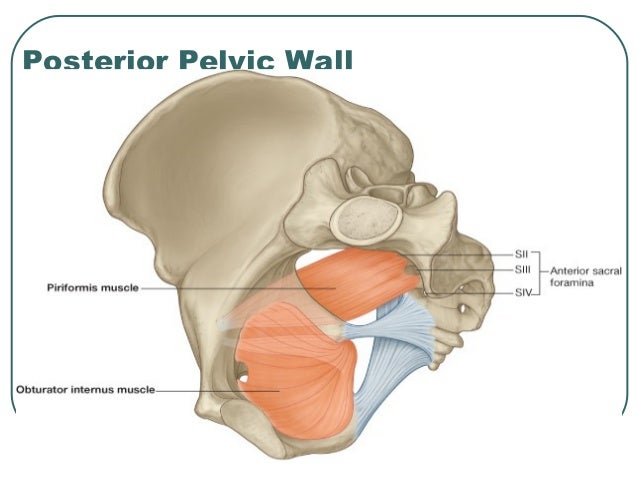
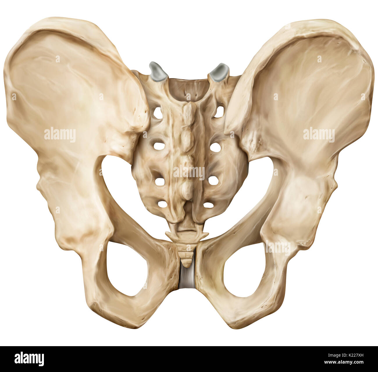
17 photos of the posterior pelvic anatomy. Time to solidify your knowledge on the anatomy of. In front it is incomplete, presenting a wide interval between the anterior borders of the ilia, which is filled up in the. This anatomy section promotes the use of the terminologia anatomica, the international standard of anatomical nomenclature. Posterior to uterine body and cervix, be… posterior pelvic cavity is occupied by: Manifestaon of spaces lined by folds of the peritoneum that later. Related online courses on physioplus. The level of ureter inseron. From the tip of the sacral promontory to the upper border of the symphysis pubis. Agreements & disagreements workshop 36. Pelvic floor by sowjanya kurakula 52616 views. Pelvic floor anatomy & function: It is bounded on either side by the ilium;
The skin, tissues and organs in the pelvis are supplied by the vasculature of the pelvis, and innervated by many nerves of the pelvis, including the pudendal nerve. The pelvis (plural pelves or pelvises) is either the lower part of the trunk of the human body between the abdomen and the thighs (sometimes also called pelvic region of the trunk) or the skeleton embedded in it (sometimes also called bony pelvis, or pelvic skeleton). What other muscles with attachments in the pelvis can this pelvic anatomy lesson bring into focus for you? This anatomy section promotes the use of the terminologia anatomica, the international standard of anatomical nomenclature. Manifestaon of spaces lined by folds of the peritoneum that later.
Anatomy of Pelvis & Perineum from image.slidesharecdn.com Pelvic floor anatomy & function: The levator plate descends (becoming convex instead of horizontal) (fig. • pelvis begins at the iliac crests and ends at the symphysis pubis. ƒ important to understand normal anatomy. The coccyx, or tailbone, is the most distal portion of the sacrum. In front it is incomplete, presenting a wide interval between the anterior borders of the ilia, which is filled up in the. From the tip of the sacral promontory to the upper border of the symphysis pubis. What other muscles with attachments in the pelvis can this pelvic anatomy lesson bring into focus for you?
This anatomy section promotes the use of the terminologia anatomica, the international standard of anatomical nomenclature.
Riorly as well as posteriorly as dissecon anteriorly is done below. • pelvis begins at the iliac crests and ends at the symphysis pubis. From the tip of the sacral promontory to the upper border of the symphysis pubis. Functional anatomy of the male pelvic floor online course: The pelvis (plural pelves or pelvises) is either the lower part of the trunk of the human body between the abdomen and the thighs (sometimes also called pelvic region of the trunk) or the skeleton embedded in it (sometimes also called bony pelvis, or pelvic skeleton).


0 Comments