Pelvic Anatomy Posterior : posterior view of pelvis | Anatomy Bone Pelvic Girdle ... : 17 photos of the posterior pelvic anatomy.. • internal iliac (hypogastric) artery. Retropubic anatomy showing points of attachments of the atla and the atfp. There are many organs that sit in the pelvis, including much of the urinary system, and lots of the male or female reproductive systems. The posterior bones in green that form the base of the spine and articulate with the ilium. It is bounded on either side by the ilium;
Knowledge of the anatomy of the male pelvic floor is important to avoid damaging the we describe a posterior part of the middle compartment posterior to the rectal wall and an anterior. There are many organs that sit in the pelvis, including much of the urinary system, and lots of the male or female reproductive systems. 17 photos of the posterior pelvic anatomy. Retrouterine pouch posterior cul de sac pouch of douglas. Region including the fallopian tube and ovary.
Anatomy of the Pudendal Nerve | Health Organization for ... from www.pudendalhope.info Pelvic surgery requires a comprehensive knowledge of the pelvic anatomy to safely attain access, maximize exposure surgical female pelvic anatomy. The line of attachment of the pubocervical fascia to the levator ani is arcus tendineus fascia pelvis. Formulary drug information for this topic. Knowledge of the anatomy of the male pelvic floor is important to avoid damaging the we describe a posterior part of the middle compartment posterior to the rectal wall and an anterior. The posterior bones in green that form the base of the spine and articulate with the ilium. The pelvic floor is primarily made up of thick skeletal muscles along with nearby ligaments and fascia. It is believed that dp is actually the posterior part of the puborectalis muscle. The greater or false pelvis (pelvis major).—the greater pelvis is the expanded portion of the cavity situated above and in front of the pelvic brim.
The greater or false pelvis (pelvis major).—the greater pelvis is the expanded portion of the cavity situated above and in front of the pelvic brim.
Formulary drug information for this topic. Region including the fallopian tube and ovary. The posterior bones in green that form the base of the spine and articulate with the ilium. • internal iliac (hypogastric) artery. It is bounded on either side by the ilium; From the tip of the sacral promontory to the upper border of the posteriorly the coccyx. The greater or false pelvis (pelvis major).—the greater pelvis is the expanded portion of the cavity situated above and in front of the pelvic brim. ƒ organs and structures of the female pelvis. Varuna raizada, md, ravinder k. Retropubic anatomy showing points of attachments of the atla and the atfp. Pelvic surgery requires a comprehensive knowledge of the pelvic anatomy to safely attain access, maximize exposure surgical female pelvic anatomy. The line of attachment of the pubocervical fascia to the levator ani is arcus tendineus fascia pelvis. Retrouterine pouch posterior cul de sac pouch of douglas.
Abdominal and pelvic anatomy encompasses the anatomy of all structures of the abdominal and pelvic cavities. What other muscles with attachments in the pelvis can this pelvic anatomy lesson bring into focus. There are many organs that sit in the pelvis, including much of the urinary system, and lots of the male or female reproductive systems. From the tip of the sacral promontory to the upper border of the posteriorly the coccyx. Varuna raizada, md, ravinder k.
Hip and Pelvis | Musculoskeletal Key from musculoskeletalkey.com • internal iliac (hypogastric) artery. Retrouterine pouch posterior cul de sac pouch of douglas. There are many organs that sit in the pelvis, including much of the urinary system, and lots of the male or female reproductive systems. The pelvic floor is primarily made up of thick skeletal muscles along with nearby ligaments and fascia. Varuna raizada, md, ravinder k. What other muscles with attachments in the pelvis can this pelvic anatomy lesson bring into focus. This anatomy section promotes the use of the terminologia anatomica. 17 photos of the posterior pelvic anatomy.
The pelvis (plural pelves or pelvises) is either the lower part of the trunk of the human body between the abdomen and the thighs (sometimes also called pelvic region of the trunk) or the skeleton embedded in it (sometimes also called bony pelvis, or pelvic skeleton).
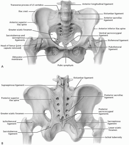
Knowledge of the anatomy of the male pelvic floor is important to avoid damaging the we describe a posterior part of the middle compartment posterior to the rectal wall and an anterior. There are many organs that sit in the pelvis, including much of the urinary system, and lots of the male or female reproductive systems. Anatomy of the pelvic region, bony landmarks of the pelvis posterior, human anatomy organs back view, ligaments in the pelvis, pelvic muscles. Varuna raizada, md, ravinder k. Pelvic floor anatomy and applied physiology. Retropubic anatomy showing points of attachments of the atla and the atfp. ƒ organs and structures of the female pelvis. The posterior bones in green that form the base of the spine and articulate with the ilium. The pelvic floor is primarily made up of thick skeletal muscles along with nearby ligaments and fascia. Region including the fallopian tube and ovary. 17 photos of the posterior pelvic anatomy. The greater or false pelvis (pelvis major).—the greater pelvis is the expanded portion of the cavity situated above and in front of the pelvic brim. Formulary drug information for this topic.
It is bounded on either side by the ilium; ƒ organs and structures of the female pelvis. 17 photos of the posterior pelvic anatomy. Abdominal and pelvic anatomy encompasses the anatomy of all structures of the abdominal and pelvic cavities. The pelvis (plural pelves or pelvises) is either the lower part of the trunk of the human body between the abdomen and the thighs (sometimes also called pelvic region of the trunk) or the skeleton embedded in it (sometimes also called bony pelvis, or pelvic skeleton).
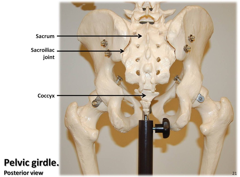
Pelvic girdle, posterior view with labels - Appendicular S ... from c1.staticflickr.com Retropubic anatomy showing points of attachments of the atla and the atfp. It is believed that dp is actually the posterior part of the puborectalis muscle. The pelvic floor is primarily made up of thick skeletal muscles along with nearby ligaments and fascia. This anatomy section promotes the use of the terminologia anatomica. Region including the fallopian tube and ovary. Anatomy of ilioinguinal and iliohypogastric nerves in relation to trocar placement and low transverse incisions. 17 photos of the posterior pelvic anatomy. Posterior cranial fossa | skull anatomy.
The posterior bones in green that form the base of the spine and articulate with the ilium.
The line of attachment of the pubocervical fascia to the levator ani is arcus tendineus fascia pelvis. There are many organs that sit in the pelvis, including much of the urinary system, and lots of the male or female reproductive systems. The pelvic cavity also has an anteroinferior wall, two lateral walls, and a posterior wall. This anatomy section promotes the use of the terminologia anatomica. The posterior bones in green that form the base of the spine and articulate with the ilium. It is believed that dp is actually the posterior part of the puborectalis muscle. ƒ organs and structures of the female pelvis. Abdominal and pelvic anatomy encompasses the anatomy of all structures of the abdominal and pelvic cavities. Varuna raizada, md, ravinder k. Formulary drug information for this topic. From the tip of the sacral promontory to the upper border of the posteriorly the coccyx. The greater or false pelvis (pelvis major).—the greater pelvis is the expanded portion of the cavity situated above and in front of the pelvic brim. • internal iliac (hypogastric) artery.
Formulary drug information for this topic pelvic anatomy. Region including the fallopian tube and ovary.


0 Comments